Human respiratory system
PHYSIOLOGY
PHYSIOLOGY

HUMAN RESPIRATORY SYSTEM
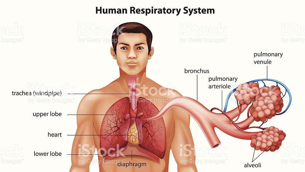
The human respiratory system is made up of many organs such as nose, nasal cavity, pharynx, larynx, trachea, bronchi and a pair of lungs. But, lungs are the main respiratory organs where exchange of gases takes place. Following is the detail of human respiratory system.
The human respiratory system is made up of many organs such as nose, nasal cavity, pharynx, larynx, trachea, bronchi and a pair of lungs. But, lungs are the main respiratory organs where exchange of gases takes place. Following is the detail of human respiratory system.
1. Nasal cavity
In human beings air containing oxygen enter the body through nose. There are present two holes in our nose called nostrils. The nostrils opens behind in the nasal cavity. The nasal cavity lies above the buccal cavity. The function of nasal cavity is to moisten, warm and trap dust particles present in the air coming in it through nostrils.The nasal cavity opens behind in the pharynx.
2. Pharynx
Pharynx is a common passage for both the respiratory and digestive system. It leads into the larynx (voice box).
3. Larynx
It is also known as voice box. It lies at the top of trachea. The pharynx opens into the larynx through a slit like opening called glottis. The glottis is protected by a leaf shaped flap of cartilage called epiglottis. Epiglottis covers the mouth of glottis when we swallow food, so that food may not enter the trachea.
There is present a pair of vocal cordsinside the larynx. When air passes through the larynx, vibrations are produced in the vocal cords and it results in production of sound.
4. Trachea
It is also known as wind pipe. It is 10-12 cm. long tube with a diameter of 2-3 cm. trachea is supported by 15-20 cartilaginous rings. These rings prevent trachea from collapsing when no air is present in it and also provides flexibility and strength.
At its lowerend the trachea divides into two smaller tubes called bronchi (singular bronchus)which enters the right and left lungs. The walls of bronchi are also supported by cartilaginous rings. In the lungs, each bronchus further divides into still smaller tubes called bronchioles. At the ends of each bronchiole there is present a cluster of tiny and thin walled sacs called alveoli. Each alveoli is surrounded by a network of very fine blood capillaries.

5. Lungs
In humans, a pair of lungs is present in the thoracic cavity, one on each side of the heart. These are called right lung and left lung respectively. The right lung is larger than the left lung. The right lung has three lobes and two fissures. While, the left lung has two lobes and only one fissure. Both the lungs are covered by two membranes called pleura. The fluid present in between these two membranes is called pleural fluid. It protects the lungs from mechanical injuries.
In humans, a pair of lungs is present in the thoracic cavity, one on each side of the heart. These are called right lung and left lung respectively. The right lung is larger than the left lung. The right lung has three lobes and two fissures. While, the left lung has two lobes and only one fissure. Both the lungs are covered by two membranes called pleura. The fluid present in between these two membranes is called pleural fluid. It protects the lungs from mechanical injuries.
Human respiratory system, the system in humans that takes up oxygen and expels carbon dioxide.

Morphology of the upper airways
The nose
The nose is the external protuberance of an internal space, the nasal cavity. It is subdivided into a left and right canal by a thin medial cartilaginous and bony wall, the nasal septum.
The nose is the external protuberance of an internal space, the nasal cavity. It is subdivided into a left and right canal by a thin medial cartilaginous and bony wall, the nasal septum.
The Human Respiratory System
The Pathway :
Air enters the nostrilspasses through the nasopharynx,the oral pharynxthrough the glottisinto the tracheainto the right and left bronchi, which branches and rebranches intobronchioles, each of which terminates in a cluster ofalveoli
Only in the alveoli does actual gas exchange takes place. There are some 300 million alveoli in two adult lungs. These provide a surface area of some 160 m2(almost equal to the singles area of a tennis court and 80 times the area of our skin!).
Breathing
In mammals, the diaphragm divides the body cavity into theabdominal cavity, which contains the viscera (e.g., stomach and intestines) and thethoracic cavity, which contains the heart and lungs.
In mammals, the diaphragm divides the body cavity into theabdominal cavity, which contains the viscera (e.g., stomach and intestines) and thethoracic cavity, which contains the heart and lungs.
The inner surface of the thoracic cavity and the outer surface of the lungs are lined with pleural membranes which adhere to each other. If air is introduced between them, the adhesion is broken and the natural elasticity of the lung causes it to collapse. This can occur from trauma. And it is sometimes induced deliberately to allow the lung to rest. In either case, reinflation occurs as the air is gradually absorbed by the tissues.
Because of this adhesion, any action that increases the volume of the thoracic cavity causes the lungs to expand, drawing air into them.During inspiration (inhaling),The external intercostal muscles contract, lifting the ribs up and out.The diaphragm contracts, drawing it down .During expiration (exhaling), these processes are reversed and the natural elasticity of the lungs returns them to their normal volume. At rest, we breath 15–18 times a minute exchanging about 500 ml of air.In more vigorous expiration,The internal intercostal muscles draw the ribs down and inwardThe wall of the abdomen contracts pushing the stomach and liver upward.Under these conditions, an average adult male can flush his lungs with about 4 liters of air at each breath. This is called the vital capacity. Even with maximum expiration, about 1200 ml of residual airremain.
The table shows what happens to the composition of air when it reaches the alveoli. Some of the oxygen dissolves in the film of moisture covering the epithelium of the alveoli. From here it diffuses into the blood in a nearby capillary. It enters a red blood cell and combines with the hemoglobin therein.
At the same time, some of the carbon dioxide in the blood diffuses into the alveoli from which it can be exhaled
Composition of atmospheric air and expired air in a typical subject.
Note that only a fraction of the oxygen inhaled is taken up by the lungs.ComponentAtmospheric Air (%)Expired Air (%)N2 (plus inert gases)78.6274.9O220.8515.3CO20.033.6H2O0.56.2 100.0%100.0%
The ease with which oxygen and carbon dioxide can pass between air and blood is clear from this electron micrograph of two alveoli (Air) and an adjacent capillary from the lung of a laboratory mouse. Note the thinness of the epithelial cells (EP) that line the alveoli and capillary (except where the nucleus is located). At the closest point, the surface of the red blood cell is only 0.7 µm away from the air in the alveolus.
Central Control of Breathing
The rate of cellular respiration (and hence oxygen consumption and carbon dioxide production) varies with level of activity. Vigorous exercise can increase by 20–25 times the demand of the tissues for oxygen. This is met by increasing the rate and depth of breathing.
It is a rising concentration of carbon dioxide — not a declining concentration of oxygen — that plays the major role in regulating the ventilation of the lungs. Certain cells in the medulla oblongata are very sensitive to a drop in pH. As the CO2content of the blood rises above normal levels, the pH drops
[CO2 + H2O → HCO3− + H+],
and the medulla oblongata responds by increasing the number and rate of nerve impulses that control the action of the intercostal muscles and diaphragm. This produces an increase in the rate of lung ventilation, which quickly brings the CO2concentration of the alveolar air, and then of the blood, back to normal levels.
However, the carotid body in the carotid arteries does have receptors that respond to a drop in oxygen. Their activation is important in situations (e.g., at high altitude in the unpressurized cabin of an aircraft) where oxygen supply is inadequate but there has been no increase in the production of CO2. People who live at high altitudes, e.g., in the Andes, have enlarged carotid bodies.

Local Control of Breathing
The smooth muscle in the walls of the bronchioles is very sensitive to the concentration of carbon dioxide. A rising level of CO2 causes the bronchioles to dilate. This lowers the resistance in the airways and thus increases the flow of air in and out.
Diseases of the Lungs
Pneumonia
Pneumonia is an infection of the alveoli. It can be caused by many kinds of both bacteria (e.g., Streptococcus pneumoniae) and viruses. Tissue fluids accumulate in the alveoli reducing the surface area exposed to air. If enough alveoli are affected, the patient may need supplemental oxygen.
Asthma
In asthma, periodic constriction of the bronchi and bronchioles makes it more difficult to breathe in and, especially, out. Attacks of asthma can betriggered by airborne irritants such as chemical fumes and cigarette smokeairborne particles to which the patient is allergic.Link to discussion of allergic asthma.

Emphysema
In this disorder, the delicate walls of the alveoli break down, reducing the gas-exchange area of the lungs. The condition develops slowly and is seldom a direct cause of death. However, the gradual loss of gas-exchange area forces the heart to pump ever-larger volumes of blood to the lungs in order to satisfy the body's needs. The added strain can lead to heart failure.
The immediate cause of emphysema seems to be the release of proteolytic enzymes as part of the inflammatory process that follows irritation of the lungs. Most people avoid this kind of damage during infections, etc. by producing an enzyme inhibitor (a serpin) called alpha-1 antitrypsin. Those rare people who inherit two defective genes for alpha-1 antitrypsin are particularly susceptible to developing emphysema.

Chronic Bronchitis
Any irritant reaching the bronchi and bronchioles will stimulate an increased secretion of mucus. In chronic bronchitis the air passages become clogged with mucus, and this leads to a persistent cough. Chronic bronchitis is usually associated with cigarette smoking.

Chronic Obstructive Pulmonary Disease (COPD)

Irritation of the lungs can lead to asthma, emphysema, and chronic bronchitis. And, in fact, many people develop two or three of these together. This constellation is known as chronic obstructive pulmonary disease (COPD).
Among the causes of COPD arecigarette smoke (often)cystic fibrosis (rare)
Cystic fibrosis is a genetic disorder caused by inheriting two defective genes for the cystic fibrosis transmembrane conductance regulator (CFTR), a transmembrane protein needed for the transport of Cl− and HCO3− ions through the plasma membrane of epithelial cells. In healthy lungs, secretion of Cl− (along with Na+ through a different channel) draws water by osmosis into the fluid bathing the cells. In cystic fibrosis, inadequate secretion of Cl− and the resulting lack of water produces a thick, sticky mucus covering the epithelia and hampering the ability of ciliated cells to move it up out of the lungs. In addition, diminished secretion of HCO3− lowers the pH of this liquid making it more hospitable to colonization by inhaled bacteria. The resulting inflammation increases the accumulation of mucus which plugs the airways interfering with breathing and causing a persistent cough. Cystic fibrosis is the most common inherited disease in the U.S. white population.
Lung Cancer :

Lung cancer is the most common cancer and the most common cause of cancer deaths in U.S. males. Although more women develop breast cancer than lung cancer, since 1987 U.S. women have been dying in larger numbers from lung cancer than from breast cancer.
Lung cancer, like all cancer, is an uncontrolled proliferation of cells. There are several forms of lung cancer, but the most common (and most rapidly increasing) types are those involving the epithelial cells lining the bronchi and bronchioles.
Ordinarily, the lining of these airways consists of two layers of cells. Chronic exposure to irritants
causes the number of layers to increase. This is especially apt to happen at forks where the bronchioles branch.The ciliated and mucus-secreting cells disappear and are replaced by a disorganized mass of cells with abnormal nuclei.If the process continues, the growing mass penetrates the underlying basement membrane.Link to illustrations of the cellular changes in developing lung cancer.At this point, malignant cells can break away and be carried in lymph and blood to other parts of the body where they may lodge and continue to proliferate.It is this metastasis of the primary tumor that eventually kills the patient.

Comments
Post a Comment
Please do not Enter any Spam Link In Comment Box